This page contains information about recent cases, the symptoms, and diagnosis. We post these on our FaceBook page and give clients the opportunity to research the symtoms and try to diagnose the case themselves before we post our diagnosis.
August 2020
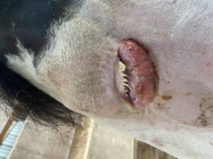
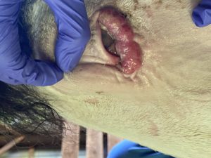
Case: You recently rescued a teenage Paint mare that has an eye that looks like this. You notice it has a foul odor, yellow discharge, and appears itchy and uncomfortable. You are also not sure she can see with this eye. Her other eye, which is blue, is completely normal and visual. While examining the rest of her face, you notice she has a sunburnt nose, but is otherwise normal with no nasal discharge.
What is your diagnosis?
Our diagnosis: Squamous Cell Carcinoma
Squamous cell carcinoma (SCC) is one of the most commonly diagnosed skin tumors in horses. Most occur at a mucocutaneous junction (where haired skin and mucous membranes merge) and are locally invasive (grow to surrounding tissues) and slow to metastasize (spread). Because of this behavior, it is important to address treatment early before they get too large. The most common locations for these tumors are the periorbital and genital regions, with the eyelid most commonly affected. Factors that increase the horse’s risk of development include chronic exposure to ultraviolet light (especially in horses with pink skin/eyelids like this Paint mare) and chronic skin irritation. There are a few different options for therapy so it is important to discuss treatment plans with your veterinarian and decide which would be best for your horse.
July 2013
Cow: 4 year old, commercial beef cow.
Case: Client X called the emergency line at 9:15 am to ask for help with a dystocia (difficult birth). One of his cows was in labor and seemed unable to deliver the calf on her own. After placing the call the farmer caught the cow and restrained her in preparation for the vet’s arrival.
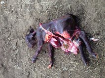
When the doctor arrived an exam was performed. The calf’s joints were very difficult to manipulate. The veterinarian was unable to correct the calf’s positioning due to the lack of flexibility. Therefore, a C-section was performed and the pictured dead fetus was removed from the cow. What is the fetal abnormality?
What is your diagnosis?
Scarlett Mobile has made it a goal this year to present a case each month in an attempt to better prepare large animal owners for cases we commonly see in our area. If you would like to submit your diagnosis for the case of the month please contact us by E-mail (scarlettmobile@hotmail.com). The doctor submitting the case will then select a person from those who give the correct answer for a free good from Scarlett Mobile.
Please submit answers by July 16th, 2013. Check back here after July 16th to read our diagnosis and treatment.
Our Diagnosis: Schistosomas Reflexus is the name for this particular fetal abnormality in cattle. Calves which develop this condition are characterized by a failure of the body wall to close, resulting in a calf that is essentially “inside-out” with exposure of theabdominal organs. The skeleton is commonly malformed, with anomalies such as decreased number or fused ribs and vertebrae, crooked and improperly positioned limbs, and inversion of the spinal canal. Any number of gross defects have been described and are highly variable. The calf may be carried to term, and the first sign of a problem may be a dystocia (difficulty giving birth) in which the cow fails to make any progress towards delivery. There is currently no known genetic correlation with this fetal abnormality. The cow recovered nicely and is doing well.
 April 2013
Derby: 15 year old, bay, Thoroughbred gelding.
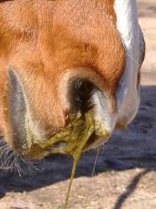
Case: Client X called the emergency line at 6:00 pm. When she got home from work and checked on the horses Derby was in distress. Her husband had fed the evening grain 1 hour earlier. Derby was not eating and had most of his grain left still. She noticed greenish colored nasal discharge combined with Derby routinely extending his neck and coughing. This routine continued at regular intervals. She called our emergency line. The on call doctor instructed her to put a halter and lead on Derby and to with hold hay, pasture and grain until the they arrived.
When the doctor arrived a physical exam was performed. Derby’s heart rate was 56 beats per minute, his respiration was 40 breathes per minute with a temperature of 101.9 F. Derby’s nasal discharge continued and began showing feed particles as well. His mucous membranes were pink with a capillary refill time (CRT) of 1 1/2 seconds. The respiratory distress and posturing while coughing continued.
What is your diagnosis?
Scarlett Mobile has made it a goal this year to present at least one case each month in an attempt to better prepare large animal owners for cases we commonly see in our area. If you would like to submit your diagnosis for the case of the month please contact us by E-mail (scarlettmobile@hotmail.com). The doctor submitting the case will then select a person from those who give the correct answer for a free good from Scarlett Mobile.
Please submit answers by April 30th, 2013. Check back here after April 30 to read our diagnosis and treatment.
Our Diagnosis: Excerpts from the Life Learn Article “Choke in Horses” You can read the full article by visiting our Life Learn Library.
Choke is the answer to the case of the month. Choke is a relatively common condition that occurs when food or a foreign body blocks the horse’s esophagus (gullet), which is the tube that takes food from the back of the mouth (pharynx) to the stomach. Choke may be partial or complete.
What causes choke?
The most common cause of choke is swallowing food or other material, that is either too dry or coarse (most commonly hay), or that swells rapidly once chewed (typically sugar beet) so that its passage down the esophagus is slowed or stopped. It can occur if a greedy horse attempts to swallow hay without chewing it thoroughly or in foals who are given access to dry, coarse hay or straw. Any condition that interferes with the horse’s ability to swallow (e.g., sedation, trauma (injury) to the neck or esophagus, grass sickness, botulism, etc.) can predispose to choke.
What are the signs of choke in horses?
The most obvious signs are discharge of saliva and feed material from the nostrils and/or mouth, depression and apparent difficulty in swallowing. When first ‘choked’ some horses will panic, make repeated unsuccessful efforts to swallow, cough and ‘gag’ as though trying to clear something from the back of the throat. If the condition has gone unnoticed, the horse may become dehydrated and severely depressed. If the esophagus ruptures, death may follow due to shock and infection. Fortunately, this is not common. Although many cases clear on their own, if you think your horse has choke, call your veterinarian immediately. The sooner treatment is applied, the sooner the condition will resolve and second complications are less likely.
How is choke treated?
In most cases, saliva continually produced in the mouth lubricates the offending obstruction, eventually allowing its passage to the stomach. Your veterinarian can help speed resolution by administering a sedative or a spasmolytic (‘antispasm’) injection to help relax the muscles of the esophagus. Sometimes, this is all that is required.
In other cases the obstruction can be gently encouraged to move on down into the stomach with the help of the stomach tube. This must be done with great care to avoid injury to the esophagus. If this cannot be achieved easily, the horse is sedated and the obstruction is flushed with water and lubricant via the stomach tube, with the head positioned lower than the esophagus. Fluid is gently pumped in via the stomach tube and allowed to run out the nostrils, gradually flushing some of the obstructing material out. This can be a long process and patience is needed to avoid damaging the esophagus. In some panic-stricken, uncooperative or solidly-obstructed cases it is necessary to anesthetize the horse to allow flushing to be performed safely and thoroughly. Once the choke is cleared the horse should be fed sloppy feeds or grass for several days to allow any local swelling to subside.
Can I prevent choke?
The most important management considerations are:
- Soak dried foodstuffs thoroughly to allow them to swell before they are eaten and swallowed.
- Ask your veterinarian to provide regular routine dental care to allow the horse to chew food thoroughly and effectively before it is swallowed. Injuries to the insides of the cheeks, caused by sharp teeth, will cause discomfort and may discourage a horse from chewing food properly.
- Provide permanent access to clean water to encourage the horse to drink normally.
- Some horses choke on a particular feed and once this is recognized, access should obviously be avoided.
Deidre M. Carson, BVSc, MRCVS & Sidney W. Ricketts, LVO, BSc, BVSc, DESM, DipECEIM, FRCPath, FRCVS.
Edited by Kim McGurrin BSc DVM DVSc Diplomate ACVIM © Copyright 2010 Lifelearn Inc. Used and/or modified with permission under license.
For more information you can view articles on our website at www.scarlettmobilevet.com under Client Education, Life Learn Library or visit the American Association of Equine Practitioners website at http://www.aaep.org/index.php
 January 2013
Blaze: 7 year old, chestnut, Quarter Horse gelding.
Case: Client X called the emergency line at 7:00 pm. Because when she went to feed Blaze his evening grain he was not interested in eating. She watched him for 10 -15 minutes while picking stalls and noticed him looking back at his sides. She was concerned that he was not eating his grain so she called our emergency line. Blaze then attempted to lay down while she was talking to the on call doctor. The on call doctor instructed her to put a halter and lead on Blaze and walk him until the they arrived.
When the doctor arrived a physical exam was performed. Blaze’s heart rate was 60 beats per minute, his respiration was 18 breathes per minute with a temperature of 100.3 F. Blaze seemed uncomfortable and kept shifting his weight on his hind legs. His mucous membranes were pink with a capillary refill time (CRT) of 2 seconds. He had reduced gut sounds in all four quadrants.
What is your diagnosis?
Scarlett Mobile has made it a goal this year to present at least one case each month in an attempt to better prepare large animal owners for cases we commonly see in our area. If you would like to submit your diagnosis for the case of the month please contact us by E-mail (scarlettmobile@hotmail.com). The doctor submitting the case will then select a person from those who give the correct answer for a free good from Scarlett Mobile.
Please submit answers by February 5th, 2013. Check back here after Feb. 5 to read our diagnosis and treatment.
Our Diagnosis:
Colic is the answer to the case of the month. Colic is the most common equine emergency that we see. It can be a confusing diagnosis for some but it is important to remember that Colic is a general term that means abdominal pain. Most of the time it is related to the GI tract in horses but anything can cause them to “colic.” The most common signs of colic are restlessness, standing off in the corner by themselves, lying down, rolling, biting at their side, flaring their nose, standing parked out, looking at their side any many more. The one consistent sign is that the horse does not want to eat which as we all know is not normal for any horse. Client X did the right thing by not ignoring the signs and staying close by to monitor Blaze. When his signs were clear to the client that something was indeed wrong, she called the emergency line to get help. We will often tell our clients to walk the horse until we can arrive. This will help to keep them from lying down, encourages gut motility, and sometimes may even relieve a gas colic. It is important to walk, do not lunge or run them in a round pen.
Heart rate is our best indicator for pain, so when Blaze’s heart rate was 60 (normal being 30-40 beats per minute), the on call vet knew we were most likely dealing with a colic. Blaze’s rectal exam revealed a small impaction in his colon. He was sedated, treated with pain relievers, and a nasogastric tube was placed in order to give him a gallon of mineral oil. He was placed in a stall for the rest of the night and was not allowed anything to eat but free choice water until he started passing manure the following morning. Blaze recovered very well and has not had any episodes since.
There are many different causes for colic. Among the most common are gas, impaction, spasms from diet change, ulcers, GI tumors, sand, displacements of bowel, pregnancy, dystocia, and many others. The important thing is to recognize the signs early and to get help.
How can I help prevent colic? There are several ways to help prevent colic. Horses like to be on a consistent routine. This means you should feed the same feeds at the same time each day. Most of their nutrition should come from forages (grass or hay) and use concentrates (grains) to complement the needs of your individual horse. Horses were made to be out grazing the majority of the day and should be allowed to be outside as much as possible. Any changes in diet should be done slowly over the course of 10-14 days and not changed suddenly. This includes forages such as hay or grass. Be sure fresh, clean water is available at all times. If you have any questions about colic, do not hesitate to call the office or the emergency line if you think your horse may be showing you signs of colic.
For more information you can view articles on our website at www.scarlettmobilevet.com under Client Education, Life Learn Library or visit the American Association of Equine Practitioners website at http://www.aaep.org/index.php

